

Description
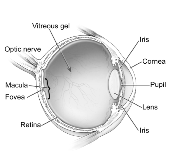
Vitelliform macular dystrophy is a genetic eye disorder that can cause worsening (progressive) vision loss. This disorder affects the retina, the specialized light-sensitive tissue that lines the back of the eye. Specifically, vitelliform macular dystrophy disrupts cells in a small area near the center of the retina called the macula. The macula is responsible for sharp central vision, which is needed for detailed tasks such as reading, driving, and recognizing faces.
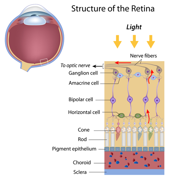

Vitelliform macular dystrophy causes a fatty yellow pigment (called lipofuscin) to build up in cells underlying the macula. Over time, large amounts of this substance can damage cells that are critical for clear central vision. As a result, people with this disorder often lose their central vision, and their eyesight may become blurry or distorted. Vitelliform macular dystrophy typically does not affect side (peripheral) vision or the ability to see at night.
Researchers have described two forms of vitelliform macular dystrophy with similar features. The early-onset form (known as Best disease) usually appears in childhood; the age at which symptoms begin and the severity of vision loss vary widely. The adult-onset form begins later, usually in mid-adulthood, and tends to cause vision loss that worsens slowly over time. The two forms of vitelliform macular dystrophy each have characteristic changes in the macula that can be detected during an eye examination.
Frequency
Vitelliform macular dystrophy is a rare disorder; its incidence is unknown.
Causes
Variants (also known as mutations) in the BEST1 and PRPH2 genes cause vitelliform macular dystrophy. BEST1 variants are responsible for Best disease and for some cases of the adult-onset form of vitelliform macular dystrophy. Changes in the PRPH2 gene can also cause the adult-onset form of vitelliform macular dystrophy; however, less than a quarter of all people with this form of the condition have variants in the BEST1 or PRPH2 gene. In most cases, the cause of the adult-onset form is unknown.
The BEST1 gene provides instructions for making a protein called bestrophin. This protein acts as a channel that controls the movement of charged chlorine atoms (chloride ions) into or out of cells in the retina. Variants in the BEST1 gene probably lead to the production of an abnormally shaped channel that cannot properly control the flow of chloride. Researchers have not determined how these malfunctioning channels are related to the buildup of lipofuscin in the macula and progressive vision loss.
The PRPH2 gene provides instructions for making a protein called peripherin 2. This protein is essential for the normal function of light-sensing (photoreceptor) cells in the retina. Variants in the PRPH2 gene cause vision loss by disrupting structures in these cells that contain light-sensing pigments. It is unclear why PRPH2 variants affect only central vision in people with adult-onset vitelliform macular dystrophy.
Inheritance
Best disease is inherited in an autosomal dominant pattern, which means one copy of the altered gene in each cell is sufficient to cause the disorder. In most cases, an affected person has one parent with the condition.

The inheritance pattern of adult-onset vitelliform macular dystrophy is uncertain. Some studies have suggested that this disorder may be inherited in an autosomal dominant pattern. It is difficult to be sure, however, because many affected people have no history of the disorder in their family, and only a small number of affected families have been reported.
Other Names for This Condition
- Vitelliform dystrophy
Additional Information & Resources
Genetic and Rare Diseases Information Center
Patient Support and Advocacy Resources
Clinical Trials
Catalog of Genes and Diseases from OMIM
Scientific Articles on PubMed
References
- Boon CJ, Klevering BJ, Leroy BP, Hoyng CB, Keunen JE, den Hollander AI. The spectrum of ocular phenotypes caused by mutations in the BEST1 gene. Prog Retin Eye Res. 2009 May;28(3):187-205. doi: 10.1016/j.preteyeres.2009.04.002. Epub 2009 Apr 16. Citation on PubMed
- Do P, Ferrucci S. Adult-onset foveomacular vitelliform dystrophy. Optometry. 2006 Apr;77(4):156-66. doi: 10.1016/j.optm.2006.01.020. Citation on PubMed
- Felbor U, Schilling H, Weber BH. Adult vitelliform macular dystrophy is frequently associated with mutations in the peripherin/RDS gene. Hum Mutat. 1997;10(4):301-9. doi: 10.1002/(SICI)1098-1004(1997)10:43.0.CO;2-J. Citation on PubMed
- Kramer F, White K, Pauleikhoff D, Gehrig A, Passmore L, Rivera A, Rudolph G, Kellner U, Andrassi M, Lorenz B, Rohrschneider K, Blankenagel A, Jurklies B, Schilling H, Schutt F, Holz FG, Weber BH. Mutations in the VMD2 gene are associated with juvenile-onset vitelliform macular dystrophy (Best disease) and adult vitelliform macular dystrophy but not age-related macular degeneration. Eur J Hum Genet. 2000 Apr;8(4):286-92. doi: 10.1038/sj.ejhg.5200447. Citation on PubMed
- MacDonald IM, Lee T, Lawrence J. Bestrophinopathies. 2003 Sep 30 [updated 2020 Jul 16]. In: Adam MP, Feldman J, Mirzaa GM, Pagon RA, Wallace SE, Bean LJH, Gripp KW, Amemiya A, editors. GeneReviews(R) [Internet]. Seattle (WA): University of Washington, Seattle; 1993-2024. Available from http://www.ncbi.nlm.nih.gov/books/NBK1167/ Citation on PubMed
- Renner AB, Tillack H, Kraus H, Kohl S, Wissinger B, Mohr N, Weber BH, Kellner U, Foerster MH. Morphology and functional characteristics in adult vitelliform macular dystrophy. Retina. 2004 Dec;24(6):929-39. doi: 10.1097/00006982-200412000-00014. Citation on PubMed
- Renner AB, Tillack H, Kraus H, Kramer F, Mohr N, Weber BH, Foerster MH, Kellner U. Late onset is common in best macular dystrophy associated with VMD2 gene mutations. Ophthalmology. 2005 Apr;112(4):586-92. doi: 10.1016/j.ophtha.2004.10.041. Citation on PubMed
- Seddon JM, Afshari MA, Sharma S, Bernstein PS, Chong S, Hutchinson A, Petrukhin K, Allikmets R. Assessment of mutations in the Best macular dystrophy (VMD2) gene in patients with adult-onset foveomacular vitelliform dystrophy, age-related maculopathy, and bull's-eye maculopathy. Ophthalmology. 2001 Nov;108(11):2060-7. doi: 10.1016/s0161-6420(01)00777-1. Citation on PubMed
- Spaide RF, Noble K, Morgan A, Freund KB. Vitelliform macular dystrophy. Ophthalmology. 2006 Aug;113(8):1392-400. doi: 10.1016/j.ophtha.2006.03.023. Citation on PubMed
- Wells J, Wroblewski J, Keen J, Inglehearn C, Jubb C, Eckstein A, Jay M, Arden G, Bhattacharya S, Fitzke F, et al. Mutations in the human retinal degeneration slow (RDS) gene can cause either retinitis pigmentosa or macular dystrophy. Nat Genet. 1993 Mar;3(3):213-8. doi: 10.1038/ng0393-213. Citation on PubMed
- White K, Marquardt A, Weber BH. VMD2 mutations in vitelliform macular dystrophy (Best disease) and other maculopathies. Hum Mutat. 2000;15(4):301-8. doi: 10.1002/(SICI)1098-1004(200004)15:43.0.CO;2-N. Citation on PubMed
The information on this site should not be used as a substitute for professional medical care or advice. Contact a health care provider if you have questions about your health.