

Description
Autosomal dominant optic atrophy and cataract is an eye disorder that is characterized by impaired vision. Most affected individuals have decreased sharpness of vision (visual acuity) from birth, while others begin to experience vision problems in early childhood or later. In affected individuals, both eyes are usually affected equally. However, the severity of the vision loss varies widely, even among affected members of the same family, ranging from nearly normal vision to complete blindness.
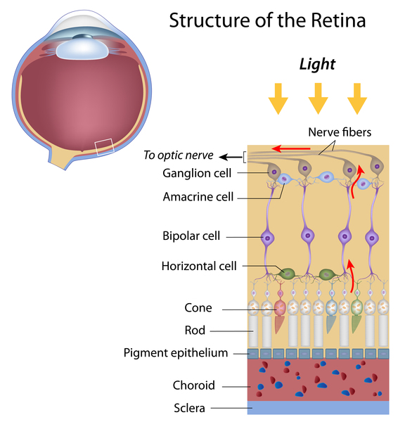
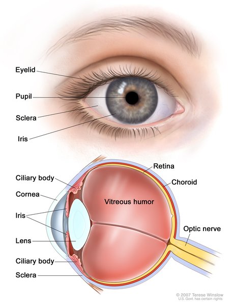
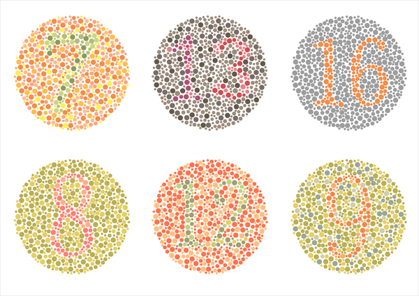
Several abnormalities contribute to impaired vision in people with autosomal dominant optic atrophy and cataract. In the early stages of the condition, affected individuals experience a progressive loss of certain cells within the retina, which is a specialized light-sensitive tissue that lines the back of the eye. The loss of these cells (known as retinal ganglion cells) is followed by the degeneration (atrophy) of the nerves that relay visual information from the eyes to the brain (optic nerves), which contributes to vision loss. Atrophy of these nerves causes an abnormally pale appearance (pallor) of the optic nerves, which can be seen only during an eye examination. Most people with this disorder also have clouding of the lenses of the eyes (cataracts). This eye abnormality can develop anytime but typically appears in childhood. Other common eye problems in autosomal dominant optic atrophy and cataract include involuntary movements of the eyes (nystagmus), or problems with color vision (color vision deficiency) that make it difficult or impossible to distinguish between shades of blue and green.
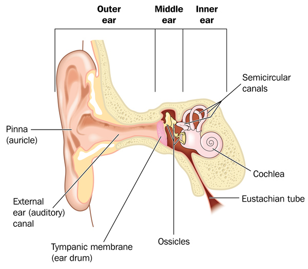
Some people with autosomal dominant optic atrophy and cataract develop disturbances in the function of other nerves (neuropathy) besides the optic nerves. These disturbances can lead to problems with balance and coordination (cerebellar ataxia), an unsteady style of walking (gait), prickling or tingling sensations (paresthesias) in the arms and legs, progressive muscle stiffness (spasticity), or rhythmic shaking (tremors). In some cases, affected individuals have hearing loss caused by abnormalities of the inner ear (sensorineural deafness).
Frequency
Autosomal dominant optic atrophy and cataract is one form of autosomal dominant optic atrophy, a group of conditions that are estimated to affect 1 in 30,000 people worldwide, and approximately 1 in 10,000 people in Denmark. A form of optic atrophy called optic atrophy type 1 accounts for most cases, while autosomal dominant optic atrophy and cataract is thought to represent only a few percent of autosomal dominant optic atrophy cases.
Causes
Autosomal dominant optic atrophy and cataract is caused by mutations in a gene called OPA3. The protein produced from this gene is made in cells and tissues throughout the body. The OPA3 protein is found within mitochondria, which are the energy-producing centers of cells. While the exact function of the protein is unknown, it is thought to play a role in the organization of the shape and structure of the mitochondria and in controlled cell death (apoptosis).

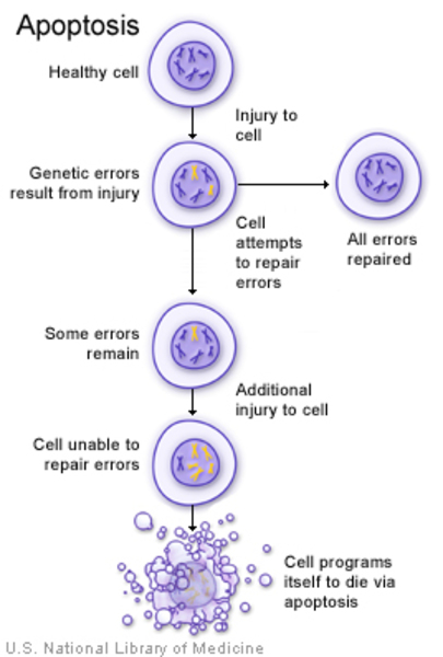
Mutations in the OPA3 gene lead to abnormal mitochondrial function. The mitochondria become misshapen and disorganized and have reduced energy-producing capabilities. Cells that contain these poorly functioning mitochondria seem to be more susceptible to apoptosis. In particular, affected cells that have high energy demands, such as retinal ganglion cells, are likely to die prematurely. Specialized extensions of retinal ganglion cells, called axons, form the optic nerves, so when retinal ganglion cells die, the optic nerves atrophy and cannot transmit visual information to the brain. Together, these effects reduce vision in affected individuals. It is likely that nerve cells in other parts of the body are similarly affected by dysfunctional mitochondria, resulting in the signs and symptoms of neuropathy in individuals with autosomal dominant optic atrophy and cataract. It is unclear how OPA3 gene mutations lead to cataracts and other eye problems that can occur in autosomal dominant optic atrophy and cataracts.

Inheritance
This condition is inherited in an autosomal dominant pattern, which means one copy of the altered gene in each cell is sufficient to cause the disorder.

In most cases, an affected person has one parent with the condition.
Other Names for This Condition
- Autosomal dominant optic atrophy type 3
- OPA3
- OPA3, autosomal dominant
- Optic atrophy and cataract, autosomal dominant
- Optic atrophy type 3
- Optic atrophy, cataract, and neurologic disorder
Additional Information & Resources
Genetic Testing Information
Genetic and Rare Diseases Information Center
Patient Support and Advocacy Resources
Catalog of Genes and Diseases from OMIM
Scientific Articles on PubMed
References
- Bagli E, Zikou AK, Agnantis N, Kitsos G. Mitochondrial Membrane Dynamics and Inherited Optic Neuropathies. In Vivo. 2017 Jul-Aug;31(4):511-525. doi: 10.21873/invivo.11090. Citation on PubMed or Free article on PubMed Central
- Grau T, Burbulla LF, Engl G, Delettre C, Delprat B, Oexle K, Leo-Kottler B, Roscioli T, Kruger R, Rapaport D, Wissinger B, Schimpf-Linzenbold S. A novel heterozygous OPA3 mutation located in the mitochondrial target sequence results in altered steady-state levels and fragmented mitochondrial network. J Med Genet. 2013 Dec;50(12):848-58. doi: 10.1136/jmedgenet-2013-101774. Epub 2013 Oct 17. Citation on PubMed
- Lenaers G, Hamel C, Delettre C, Amati-Bonneau P, Procaccio V, Bonneau D, Reynier P, Milea D. Dominant optic atrophy. Orphanet J Rare Dis. 2012 Jul 9;7:46. doi: 10.1186/1750-1172-7-46. Citation on PubMed or Free article on PubMed Central
- Li Y, Li J, Jia X, Xiao X, Li S, Guo X. Genetic and Clinical Analyses of DOA and LHON in 304 Chinese Patients with Suspected Childhood-Onset Hereditary Optic Neuropathy. PLoS One. 2017 Jan 12;12(1):e0170090. doi: 10.1371/journal.pone.0170090. eCollection 2017. Citation on PubMed or Free article on PubMed Central
- Reynier P, Amati-Bonneau P, Verny C, Olichon A, Simard G, Guichet A, Bonnemains C, Malecaze F, Malinge MC, Pelletier JB, Calvas P, Dollfus H, Belenguer P, Malthiery Y, Lenaers G, Bonneau D. OPA3 gene mutations responsible for autosomal dominant optic atrophy and cataract. J Med Genet. 2004 Sep;41(9):e110. doi: 10.1136/jmg.2003.016576. No abstract available. Citation on PubMed or Free article on PubMed Central
- Sergouniotis PI, Perveen R, Thiselton DL, Giannopoulos K, Sarros M, Davies JR, Biswas S, Ansons AM, Ashworth JL, Lloyd IC, Black GC, Votruba M. Clinical and molecular genetic findings in autosomal dominant OPA3-related optic neuropathy. Neurogenetics. 2015 Jan;16(1):69-75. doi: 10.1007/s10048-014-0416-y. Epub 2014 Aug 27. Citation on PubMed
The information on this site should not be used as a substitute for professional medical care or advice. Contact a health care provider if you have questions about your health.