

Description
Progressive external ophthalmoplegia is a condition characterized by weakness of the eye muscles. The condition typically appears in adults between ages 18 and 40 and slowly worsens over time. The first sign of progressive external ophthalmoplegia is typically drooping eyelids (ptosis), which can affect one or both eyelids. As ptosis worsens, affected individuals may use the forehead muscles to try to lift the eyelids, or they may lift up their chin in order to see. Another characteristic feature of progressive external ophthalmoplegia is weakness or paralysis of the muscles that move the eye (ophthalmoplegia). Affected individuals have to turn their head to see in different directions, especially as the ophthalmoplegia worsens. People with progressive external ophthalmoplegia may also have general weakness of the muscles used for movement (myopathy), particularly those in the neck, arms, or legs. The weakness may be especially noticeable during exercise (exercise intolerance). Muscle weakness may also cause difficulty swallowing (dysphagia).

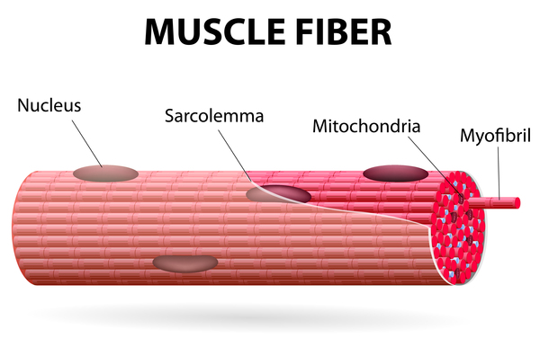
When the muscle cells of affected individuals are stained and viewed under a microscope, these cells usually appear abnormal. These abnormal muscle cells contain an excess of cell structures called mitochondria and are known as ragged-red fibers.
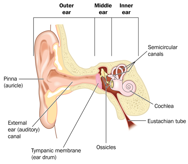
Although muscle weakness is the primary symptom of progressive external ophthalmoplegia, this condition can be accompanied by other signs and symptoms. In these instances, the condition is referred to as progressive external ophthalmoplegia plus (PEO+). Additional signs and symptoms can include hearing loss caused by nerve damage in the inner ear (sensorineural hearing loss), weakness and loss of sensation in the limbs due to nerve damage (neuropathy), impaired muscle coordination (ataxia), a pattern of movement abnormalities known as parkinsonism, and depression.
Progressive external ophthalmoplegia is part of a spectrum of disorders with overlapping signs and symptoms. Similar disorders include ataxia neuropathy spectrum and Kearns-Sayre syndrome. Like progressive external ophthalmoplegia, the other conditions in this spectrum can involve weakness of the eye muscles. However, these conditions have many additional features not shared by most people with progressive external ophthalmoplegia.
Frequency
The prevalence of progressive external ophthalmoplegia is unknown.
Causes
Progressive external ophthalmoplegia is a condition caused by defects in mitochondria, which are structures within cells that use oxygen to convert the energy from food into a form cells can use. This process is called oxidative phosphorylation. Although most DNA is packaged in chromosomes within the nucleus (nuclear DNA), mitochondria also have a small amount of their own DNA, called mitochondrial DNA or mtDNA. This DNA contains genes essential for oxidative phosphorylation.
Progressive external ophthalmoplegia can result from mutations in one of several different genes. In some cases, mutations in nuclear DNA are responsible for the condition, including mutations in the POLG, TWNK, RRM2B, and SLC25A4 genes, among others. These genes are critical for the production and maintenance of mtDNA. Although the mechanism is unclear, mutations in these genes lead to the deletion of large segments of mtDNA in muscle cells. The size of the deleted region can range from 2,000 to 10,000 DNA building blocks (nucleotides).
In other cases, the condition is caused by a single large deletion of mtDNA that is not associated with a mutation in a nuclear DNA gene.
Less commonly, mutations that change single nucleotides in genes found in mtDNA, such as the MT-TL1 gene, cause progressive external ophthalmoplegia. These mutations occur in genes that provide instructions for making molecules called transfer RNAs. Transfer RNAs help assemble protein building blocks (amino acids) into functioning proteins. The transfer RNAs associated with progressive external ophthalmoplegia are present in mitochondria and help assemble the proteins that carry out the steps of oxidative phosphorylation.
Researchers have not determined how deletions of mtDNA or mutations in mtDNA genes lead to the specific signs and symptoms of progressive external ophthalmoplegia, although the features of the condition are probably related to impaired oxidative phosphorylation. It has been suggested that eye muscles are commonly affected by mitochondrial defects because they are especially dependent on oxidative phosphorylation for energy.
Inheritance
Progressive external ophthalmoplegia can have different inheritance patterns depending on the gene involved.
When the nuclear genes POLG, TWNK, RRM2B, or SLC25A4 are involved, progressive external ophthalmoplegia is usually inherited in an autosomal dominant pattern, which means one copy of the altered gene in each cell is sufficient to cause the disorder.

Certain mutations in the POLG or RRM2B gene can also cause a form of the condition that is inherited in an autosomal recessive pattern, which means both copies of the gene in each cell have mutations. The parents of an individual with an autosomal recessive condition each carry one copy of the mutated gene, but they typically do not show signs and symptoms of the condition.

When this condition is caused by mutations in the MT-TL1 gene and other mitochondrial transfer RNA genes, it is inherited in a mitochondrial pattern, which is also known as maternal inheritance. This pattern of inheritance applies to genes contained in mtDNA. Because egg cells, but not sperm cells, contribute mitochondria to the developing embryo, children can inherit disorders resulting from mtDNA mutations only from their mother. These disorders can appear in every generation of a family and can affect both males and females, but fathers do not pass traits associated with changes in mtDNA to their children.
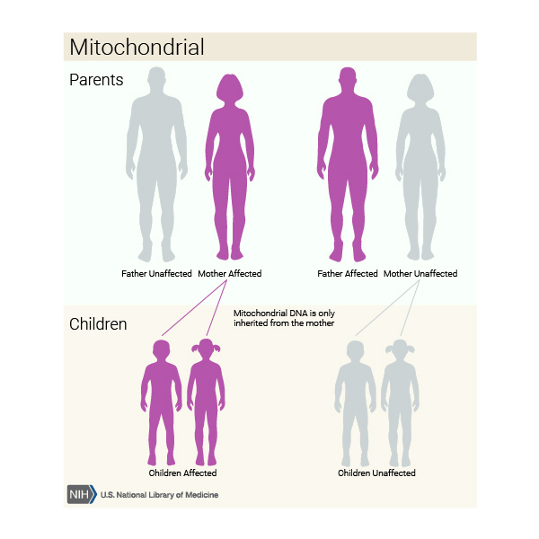
Single, large deletions of mtDNA are typically not inherited but occur during the formation of a mother's egg cells or in early development of the embryo. Individuals with these mutations usually have no history of the disorder in their family.
Other Names for This Condition
- Chronic progressive external ophthalmoplegia
- CPEO
- PEO
Additional Information & Resources
Genetic Testing Information
- Genetic Testing Registry: Progressive external ophthalmoplegia with mitochondrial DNA deletions, autosomal dominant 1

- Genetic Testing Registry: Progressive external ophthalmoplegia with mitochondrial DNA deletions, autosomal dominant 2
- Genetic Testing Registry: Progressive external ophthalmoplegia with mitochondrial DNA deletions, autosomal dominant 3
- Genetic Testing Registry: Progressive external ophthalmoplegia
Genetic and Rare Diseases Information Center
Patient Support and Advocacy Resources
Clinical Trials
Catalog of Genes and Diseases from OMIM
- PROGRESSIVE EXTERNAL OPHTHALMOPLEGIA WITH MITOCHONDRIAL DNA DELETIONS, AUTOSOMAL DOMINANT 1; PEOA1
- PROGRESSIVE EXTERNAL OPHTHALMOPLEGIA WITH MITOCHONDRIAL DNA DELETIONS, AUTOSOMAL RECESSIVE 1; PEOB1
- PROGRESSIVE EXTERNAL OPHTHALMOPLEGIA WITH MITOCHONDRIAL DNA DELETIONS, AUTOSOMAL DOMINANT 2; PEOA2
- PROGRESSIVE EXTERNAL OPHTHALMOPLEGIA WITH MITOCHONDRIAL DNA DELETIONS, AUTOSOMAL DOMINANT 3; PEOA3
Scientific Articles on PubMed
References
- Chan SS, Longley MJ, Copeland WC. The common A467T mutation in the human mitochondrial DNA polymerase (POLG) compromises catalytic efficiency and interaction with the accessory subunit. J Biol Chem. 2005 Sep 9;280(36):31341-6. doi: 10.1074/jbc.M506762200. Epub 2005 Jul 16. Citation on PubMed
- Cohen BH, Chinnery PF, Copeland WC. POLG-Related Disorders. 2010 Mar 16 [updated 2024 Feb 29]. In: Adam MP, Feldman J, Mirzaa GM, Pagon RA, Wallace SE, Bean LJH, Gripp KW, Amemiya A, editors. GeneReviews(R) [Internet]. Seattle (WA): University of Washington, Seattle; 1993-2024. Available from http://www.ncbi.nlm.nih.gov/books/NBK26471/ Citation on PubMed
- Goffart S, Cooper HM, Tyynismaa H, Wanrooij S, Suomalainen A, Spelbrink JN. Twinkle mutations associated with autosomal dominant progressive external ophthalmoplegia lead to impaired helicase function and in vivo mtDNA replication stalling. Hum Mol Genet. 2009 Jan 15;18(2):328-40. doi: 10.1093/hmg/ddn359. Epub 2008 Oct 29. Citation on PubMed or Free article on PubMed Central
- Kaukonen J, Juselius JK, Tiranti V, Kyttala A, Zeviani M, Comi GP, Keranen S, Peltonen L, Suomalainen A. Role of adenine nucleotide translocator 1 in mtDNA maintenance. Science. 2000 Aug 4;289(5480):782-5. doi: 10.1126/science.289.5480.782. Citation on PubMed
- Koga Y, Akita Y, Takane N, Sato Y, Kato H. Heterogeneous presentation in A3243G mutation in the mitochondrial tRNA(Leu(UUR)) gene. Arch Dis Child. 2000 May;82(5):407-11. doi: 10.1136/adc.82.5.407. Citation on PubMed or Free article on PubMed Central
- Milone M, Massie R. Polymerase gamma 1 mutations: clinical correlations. Neurologist. 2010 Mar;16(2):84-91. doi: 10.1097/NRL.0b013e3181c78a89. Citation on PubMed
- Pitceathly RD, Smith C, Fratter C, Alston CL, He L, Craig K, Blakely EL, Evans JC, Taylor J, Shabbir Z, Deschauer M, Pohl U, Roberts ME, Jackson MC, Halfpenny CA, Turnpenny PD, Lunt PW, Hanna MG, Schaefer AM, McFarland R, Horvath R, Chinnery PF, Turnbull DM, Poulton J, Taylor RW, Gorman GS. Adults with RRM2B-related mitochondrial disease have distinct clinical and molecular characteristics. Brain. 2012 Nov;135(Pt 11):3392-403. doi: 10.1093/brain/aws231. Epub 2012 Oct 29. Citation on PubMed or Free article on PubMed Central
- Sharer JD. The adenine nucleotide translocase type 1 (ANT1): a new factor in mitochondrial disease. IUBMB Life. 2005 Sep;57(9):607-14. doi: 10.1080/15216540500217735. Citation on PubMed
- Spelbrink JN, Li FY, Tiranti V, Nikali K, Yuan QP, Tariq M, Wanrooij S, Garrido N, Comi G, Morandi L, Santoro L, Toscano A, Fabrizi GM, Somer H, Croxen R, Beeson D, Poulton J, Suomalainen A, Jacobs HT, Zeviani M, Larsson C. Human mitochondrial DNA deletions associated with mutations in the gene encoding Twinkle, a phage T7 gene 4-like protein localized in mitochondria. Nat Genet. 2001 Jul;28(3):223-31. doi: 10.1038/90058. Erratum In: Nat Genet 2001 Sep;29(1):100. Citation on PubMed
- Stumpf JD, Copeland WC. Mitochondrial DNA replication and disease: insights from DNA polymerase gamma mutations. Cell Mol Life Sci. 2011 Jan;68(2):219-33. doi: 10.1007/s00018-010-0530-4. Epub 2010 Oct 8. Citation on PubMed or Free article on PubMed Central
- Tyynismaa H, Ylikallio E, Patel M, Molnar MJ, Haller RG, Suomalainen A. A heterozygous truncating mutation in RRM2B causes autosomal-dominant progressive external ophthalmoplegia with multiple mtDNA deletions. Am J Hum Genet. 2009 Aug;85(2):290-5. doi: 10.1016/j.ajhg.2009.07.009. Epub 2009 Aug 6. Citation on PubMed or Free article on PubMed Central
- Van Goethem G, Martin JJ, Van Broeckhoven C. Progressive external ophthalmoplegia characterized by multiple deletions of mitochondrial DNA: unraveling the pathogenesis of human mitochondrial DNA instability and the initiation of a genetic classification. Neuromolecular Med. 2003;3(3):129-46. doi: 10.1385/NMM:3:3:129. Citation on PubMed
- Yu Wai Man CY, Chinnery PF, Griffiths PG. Extraocular muscles have fundamentally distinct properties that make them selectively vulnerable to certain disorders. Neuromuscul Disord. 2005 Jan;15(1):17-23. doi: 10.1016/j.nmd.2004.10.002. Epub 2004 Nov 26. Citation on PubMed
The information on this site should not be used as a substitute for professional medical care or advice. Contact a health care provider if you have questions about your health.