

Description
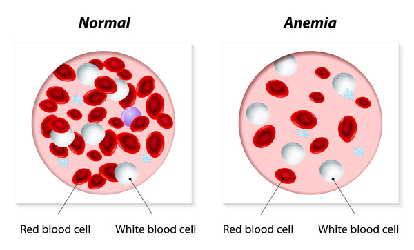
Nephronophthisis is a disorder that affects the kidneys. It is characterized by inflammation and scarring (fibrosis) that impairs kidney function. These abnormalities lead to increased urine production (polyuria), excessive thirst (polydipsia), general weakness, and extreme tiredness (fatigue). In addition, affected individuals develop fluid-filled cysts in the kidneys, usually in an area known as the corticomedullary region. Another feature of nephronophthisis is a shortage of red blood cells, a condition known as anemia.
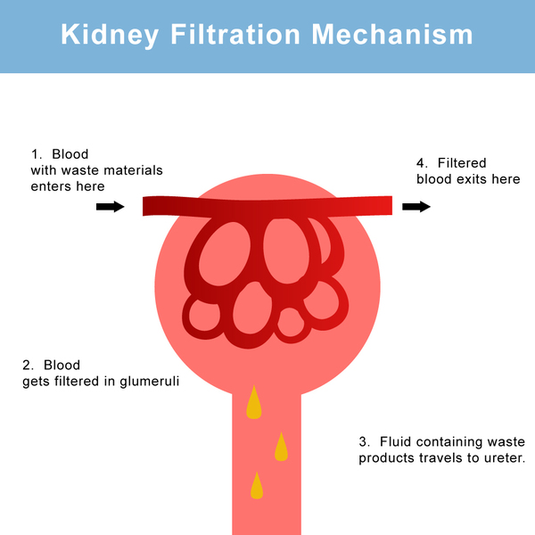
Nephronophthisis eventually leads to end-stage renal disease (ESRD), a life-threatening failure of kidney function that occurs when the kidneys are no longer able to filter fluids and waste products from the body effectively. Nephronophthisis can be classified by the approximate age at which ESRD begins: around age 1 (infantile), around age 13 (juvenile), and around age 19 (adolescent).
About 85 percent of all cases of nephronophthisis are isolated, which means they occur without other signs and symptoms. Some people with nephronophthisis have additional features, which can include liver fibrosis, heart abnormalities, or mirror image reversal of the position of one or more organs inside the body (situs inversus).
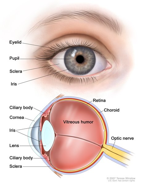
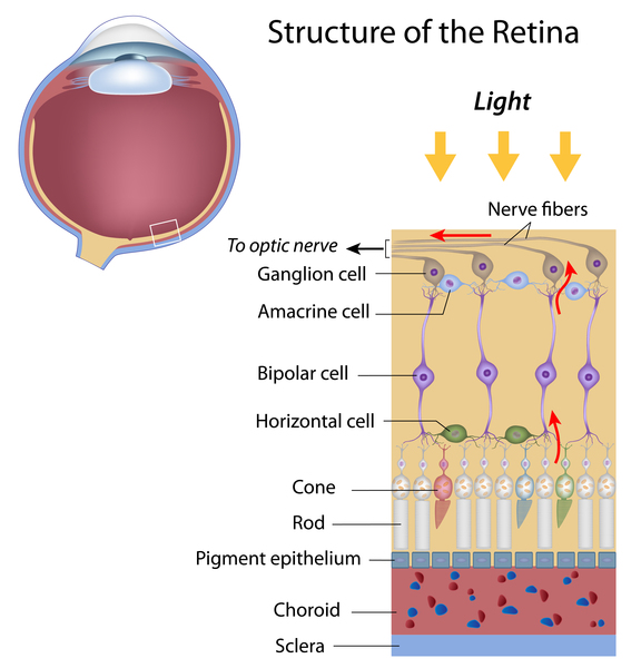
Nephronophthisis can occur as part of separate syndromes that affect other areas of the body; these are often referred to as nephronophthisis-associated ciliopathies. For example, Senior-Løken syndrome is characterized by the combination of nephronophthisis and a breakdown of the light-sensitive tissue at the back of the eye (retinal degeneration); Joubert syndrome affects many parts of the body, causing neurological problems and other features, which can include nephronophthisis.
Frequency
Nephronophthisis is found in populations worldwide. It occurs in an estimated 1 in 50,000 newborns in Canada, 1 in 100,000 in Finland, and 1 in 922,000 in the United States. Its incidence in other populations is unknown. Nephronophthisis is the most common genetic cause of ESRD in children and young adults.
Causes
Nephronophthisis has several genetic causes, which are used to split the condition into distinct types. Nephronophthisis type 1, which is the most common type of the disorder and one cause of juvenile nephronophthisis, results from changes affecting the NPHP1 gene. The proteins produced from NPHP1 and the other genes involved in nephronophthisis are known or suspected to play roles in cell structures called cilia. Cilia are microscopic, finger-like projections that stick out from the surface of cells and are involved in chemical signaling. Cilia are important for the structure and function of many types of cells and tissues, including cells in the kidneys, liver, brain, and the light-sensitive tissue at the back of the eye (the retina).
The genetic mutations involved in nephronophthisis are thought to impair the structure or function of cilia in some way, which likely disrupts important chemical signaling pathways during development. Although researchers believe that defective cilia lead to the features of nephronophthisis, the mechanism remains unclear. It is unknown why some people with mutations in nephronophthisis-associated genes have only kidney problems, while others develop additional signs and symptoms.
Inheritance
This condition is inherited in an autosomal recessive pattern, which means both copies of the gene in each cell have mutations. The parents of an individual with an autosomal recessive condition each carry one copy of the mutated gene, but they typically do not show signs and symptoms of the condition.

Other Names for This Condition
- NPH
- NPHP
Additional Information & Resources
Genetic Testing Information
- Genetic Testing Registry: Nephronophthisis 1

- Genetic Testing Registry: Infantile nephronophthisis
- Genetic Testing Registry: Nephronophthisis
- Genetic Testing Registry: Nephronophthisis 11
- Genetic Testing Registry: Nephronophthisis 12
- Genetic Testing Registry: Nephronophthisis 14
- Genetic Testing Registry: Nephronophthisis 15
- Genetic Testing Registry: Nephronophthisis 16
- Genetic Testing Registry: Nephronophthisis 18
- Genetic Testing Registry: Nephronophthisis 3
- Genetic Testing Registry: Nephronophthisis 4
- Genetic Testing Registry: Nephronophthisis 7
- Genetic Testing Registry: Nephronophthisis 9
Genetic and Rare Diseases Information Center
Patient Support and Advocacy Resources
Clinical Trials
Catalog of Genes and Diseases from OMIM
- NEPHRONOPHTHISIS 1; NPHP1
- NEPHRONOPHTHISIS 2; NPHP2
- NEPHRONOPHTHISIS 3; NPHP3
- NEPHRONOPHTHISIS 4; NPHP4
- NEPHRONOPHTHISIS 7; NPHP7
- NEPHRONOPHTHISIS 16; NPHP16
- NEPHRONOPHTHISIS 13; NPHP13
- NEPHRONOPHTHISIS 15; NPHP15
- NEPHRONOPHTHISIS 11; NPHP11
- NEPHRONOPHTHISIS 14; NPHP14
- NEPHRONOPHTHISIS 18; NPHP18
- NEPHRONOPHTHISIS 12; NPHP12
- NEPHRONOPHTHISIS 9; NPHP9
Scientific Articles on PubMed
References
- Benzing T, Schermer B. Clinical spectrum and pathogenesis of nephronophthisis. Curr Opin Nephrol Hypertens. 2012 May;21(3):272-8. doi: 10.1097/MNH.0b013e3283520f17. Citation on PubMed
- Hildebrandt F, Otto E, Rensing C, Nothwang HG, Vollmer M, Adolphs J, Hanusch H, Brandis M. A novel gene encoding an SH3 domain protein is mutated in nephronophthisis type 1. Nat Genet. 1997 Oct;17(2):149-53. doi: 10.1038/ng1097-149. Citation on PubMed
- Hurd TW, Hildebrandt F. Mechanisms of nephronophthisis and related ciliopathies. Nephron Exp Nephrol. 2011;118(1):e9-14. doi: 10.1159/000320888. Epub 2010 Nov 11. Citation on PubMed or Free article on PubMed Central
- Saunier S, Calado J, Benessy F, Silbermann F, Heilig R, Weissenbach J, Antignac C. Characterization of the NPHP1 locus: mutational mechanism involved in deletions in familial juvenile nephronophthisis. Am J Hum Genet. 2000 Mar;66(3):778-89. doi: 10.1086/302819. Citation on PubMed or Free article on PubMed Central
- Wolf MT, Hildebrandt F. Nephronophthisis. Pediatr Nephrol. 2011 Feb;26(2):181-94. doi: 10.1007/s00467-010-1585-z. Epub 2010 Jul 22. Citation on PubMed or Free article on PubMed Central
The information on this site should not be used as a substitute for professional medical care or advice. Contact a health care provider if you have questions about your health.