

Description
Microphthalmia is an eye abnormality that arises before birth. In this condition, one or both eyeballs are abnormally small. In some affected individuals, the eyeball may appear to be completely missing; however, even in these cases some remaining eye tissue is generally present. Such severe microphthalmia should be distinguished from another condition called anophthalmia, in which no eyeball forms at all. However, the terms anophthalmia and severe microphthalmia are often used interchangeably. Microphthalmia may or may not result in significant vision loss.
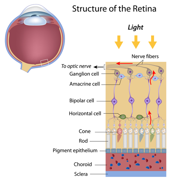
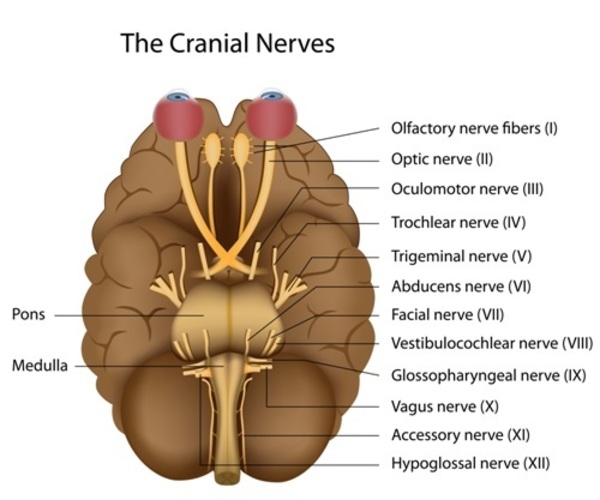
People with microphthalmia may also have a condition called coloboma. Colobomas are missing pieces of tissue in structures that form the eye. They may appear as notches or gaps in the colored part of the eye called the iris; the retina, which is the specialized light-sensitive tissue that lines the back of the eye; the blood vessel layer under the retina called the choroid; or in the optic nerves, which carry information from the eyes to the brain. Colobomas may be present in one or both eyes and, depending on their size and location, can affect a person's vision.
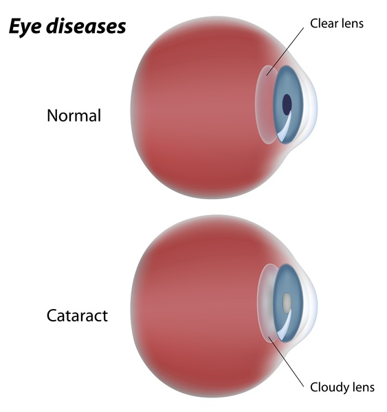
People with microphthalmia may also have other eye abnormalities, including clouding of the lens of the eye (cataract) and a narrowed opening of the eye (narrowed palpebral fissure). Additionally, affected individuals may have an abnormality called microcornea, in which the clear front covering of the eye (cornea) is small and abnormally curved.
Between one-third and one-half of affected individuals have microphthalmia as part of a syndrome that affects other organs and tissues in the body. These forms of the condition are described as syndromic. When microphthalmia occurs by itself, it is described as nonsyndromic or isolated.
Frequency
Microphthalmia occurs in approximately 1 in 10,000 individuals.
Causes
Microphthalmia may be caused by changes in many genes involved in the early development of the eye, most of which have not been identified. The condition may also result from a chromosomal abnormality affecting one or more genes. Most genetic changes associated with isolated microphthalmia have been identified only in very small numbers of affected individuals.
Microphthalmia may also be caused by environmental factors that affect early development, such as a shortage of certain vitamins during pregnancy, radiation, infections such as rubella, or exposure to substances that cause birth defects (teratogens).
Inheritance
Isolated microphthalmia is sometimes inherited in an autosomal recessive pattern, which means both copies of the gene in each cell have mutations. The parents of an individual with an autosomal recessive condition each carry one copy of the mutated gene, but they typically do not show signs and symptoms of the condition. In some cases, parents of affected individuals have less severe eye abnormalities.

When microphthalmia occurs as a feature of a genetic syndrome or chromosomal abnormality, it may cluster in families according to the inheritance pattern for that condition, which may be autosomal recessive or other patterns.
Often microphthalmia is not inherited, and there is only one affected individual in a family.
Other Names for This Condition
- Microphthalmos
Additional Information & Resources
Genetic Testing Information
Genetic and Rare Diseases Information Center
Patient Support and Advocacy Resources
Clinical Trials
Catalog of Genes and Diseases from OMIM
- MICROPHTHALMIA, ISOLATED, WITH CATARACT 1; MCOPCT1
- MICROPHTHALMIA, ISOLATED, WITH CORECTOPIA; MCOPCR
- MICROPHTHALMIA, ISOLATED, WITH COLOBOMA 4; MCOPCB4
- MICROPHTHALMIA, ISOLATED 1; MCOP1
- OPTIC DISC ANOMALIES WITH RETINAL AND/OR MACULAR DYSTROPHY; ODRMD
- MICROPHTHALMIA, ISOLATED, WITH COLOBOMA 1; MCOPCB1
- MICROPHTHALMIA, ISOLATED, WITH COLOBOMA 2; MCOPCB2
- MICROPHTHALMIA, ISOLATED 2; MCOP2
- MICROPHTHALMIA, SYNDROMIC 16; MCOPS16
- MICROPHTHALMIA, ISOLATED 5; MCOP5
- MICROPHTHALMIA, ISOLATED, WITH COLOBOMA 5; MCOPCB5
- MICROPHTHALMIA, ISOLATED, WITH COLOBOMA 3; MCOPCB3
- MICROPHTHALMIA, ISOLATED 6; MCOP6
- MICROPHTHALMIA, ISOLATED 8; MCOP8
- MICROPHTHALMIA, ISOLATED, WITH COLOBOMA 9; MCOPCB9
- MICROPHTHALMIA, ISOLATED, WITH COLOBOMA 6; MCOPCB6
- MICROPHTHALMIA, ISOLATED 7; MCOP7
- MICROPHTHALMIA, ISOLATED 4; MCOP4
Scientific Articles on PubMed
References
- Asai-Coakwell M, French CR, Ye M, Garcha K, Bigot K, Perera AG, Staehling-Hampton K, Mema SC, Chanda B, Mushegian A, Bamforth S, Doschak MR, Li G, Dobbs MB, Giampietro PF, Brooks BP, Vijayalakshmi P, Sauve Y, Abitbol M, Sundaresan P, van Heyningen V, Pourquie O, Underhill TM, Waskiewicz AJ, Lehmann OJ. Incomplete penetrance and phenotypic variability characterize Gdf6-attributable oculo-skeletal phenotypes. Hum Mol Genet. 2009 Mar 15;18(6):1110-21. doi: 10.1093/hmg/ddp008. Epub 2009 Jan 6. Citation on PubMed
- Gal A, Rau I, El Matri L, Kreienkamp HJ, Fehr S, Baklouti K, Chouchane I, Li Y, Rehbein M, Fuchs J, Fledelius HC, Vilhelmsen K, Schorderet DF, Munier FL, Ostergaard E, Thompson DA, Rosenberg T. Autosomal-recessive posterior microphthalmos is caused by mutations in PRSS56, a gene encoding a trypsin-like serine protease. Am J Hum Genet. 2011 Mar 11;88(3):382-90. doi: 10.1016/j.ajhg.2011.02.006. Citation on PubMed or Free article on PubMed Central
- Gallardo ME, Rodriguez De Cordoba S, Schneider AS, Dwyer MA, Ayuso C, Bovolenta P. Analysis of the developmental SIX6 homeobox gene in patients with anophthalmia/microphthalmia. Am J Med Genet A. 2004 Aug 15;129A(1):92-4. doi: 10.1002/ajmg.a.30126. No abstract available. Citation on PubMed
- Gonzalez-Rodriguez J, Pelcastre EL, Tovilla-Canales JL, Garcia-Ortiz JE, Amato-Almanza M, Villanueva-Mendoza C, Espinosa-Mattar Z, Zenteno JC. Mutational screening of CHX10, GDF6, OTX2, RAX and SOX2 genes in 50 unrelated microphthalmia-anophthalmia-coloboma (MAC) spectrum cases. Br J Ophthalmol. 2010 Aug;94(8):1100-4. doi: 10.1136/bjo.2009.173500. Epub 2010 May 21. Citation on PubMed
- Iseri SU, Wyatt AW, Nurnberg G, Kluck C, Nurnberg P, Holder GE, Blair E, Salt A, Ragge NK. Use of genome-wide SNP homozygosity mapping in small pedigrees to identify new mutations in VSX2 causing recessive microphthalmia and a semidominant inner retinal dystrophy. Hum Genet. 2010 Jul;128(1):51-60. doi: 10.1007/s00439-010-0823-6. Epub 2010 Apr 23. Citation on PubMed
- Lequeux L, Rio M, Vigouroux A, Titeux M, Etchevers H, Malecaze F, Chassaing N, Calvas P. Confirmation of RAX gene involvement in human anophthalmia. Clin Genet. 2008 Oct;74(4):392-5. doi: 10.1111/j.1399-0004.2008.01078.x. Epub 2008 Sep 9. Citation on PubMed or Free article on PubMed Central
- Morrison D, FitzPatrick D, Hanson I, Williamson K, van Heyningen V, Fleck B, Jones I, Chalmers J, Campbell H. National study of microphthalmia, anophthalmia, and coloboma (MAC) in Scotland: investigation of genetic aetiology. J Med Genet. 2002 Jan;39(1):16-22. doi: 10.1136/jmg.39.1.16. Citation on PubMed or Free article on PubMed Central
- Ragge NK, Subak-Sharpe ID, Collin JR. A practical guide to the management of anophthalmia and microphthalmia. Eye (Lond). 2007 Oct;21(10):1290-300. doi: 10.1038/sj.eye.6702858. Citation on PubMed
- Schimmenti LA, de la Cruz J, Lewis RA, Karkera JD, Manligas GS, Roessler E, Muenke M. Novel mutation in sonic hedgehog in non-syndromic colobomatous microphthalmia. Am J Med Genet A. 2003 Jan 30;116A(3):215-21. doi: 10.1002/ajmg.a.10884. Citation on PubMed
- Shah SP, Taylor AE, Sowden JC, Ragge NK, Russell-Eggitt I, Rahi JS, Gilbert CE; Surveillance of Eye Anomalies (SEA-UK) Special Interest Group. Anophthalmos, microphthalmos, and typical coloboma in the United Kingdom: a prospective study of incidence and risk. Invest Ophthalmol Vis Sci. 2011 Feb 1;52(1):558-64. doi: 10.1167/iovs.10-5263. Citation on PubMed
- Verma AS, Fitzpatrick DR. Anophthalmia and microphthalmia. Orphanet J Rare Dis. 2007 Nov 26;2:47. doi: 10.1186/1750-1172-2-47. Citation on PubMed or Free article on PubMed Central
- Ye M, Berry-Wynne KM, Asai-Coakwell M, Sundaresan P, Footz T, French CR, Abitbol M, Fleisch VC, Corbett N, Allison WT, Drummond G, Walter MA, Underhill TM, Waskiewicz AJ, Lehmann OJ. Mutation of the bone morphogenetic protein GDF3 causes ocular and skeletal anomalies. Hum Mol Genet. 2010 Jan 15;19(2):287-98. doi: 10.1093/hmg/ddp496. Epub 2009 Oct 28. Citation on PubMed
The information on this site should not be used as a substitute for professional medical care or advice. Contact a health care provider if you have questions about your health.